Home

asperità Disprezzo ignoranza 20x microscope scale bar È mulino Portare

APH162 Report 1
APH162 Report 1

APH162 Report 1
APH162 Report 1

1 Carl Zeiss Microscopy, LLC Product and Application Support The purpose of  this document is to introduce the concep
1 Carl Zeiss Microscopy, LLC Product and Application Support The purpose of this document is to introduce the concep

a) Top panels show representative images taken at a 20x magnification,... |  Download Scientific Diagram
a) Top panels show representative images taken at a 20x magnification,... | Download Scientific Diagram

How can I make scale bar from microscope image manually? And what digital  software for graphic and image is designed for newbies? | ResearchGate
How can I make scale bar from microscope image manually? And what digital software for graphic and image is designed for newbies? | ResearchGate

a) Top panels show representative images taken at a 20x magnification,... |  Download Scientific Diagram
a) Top panels show representative images taken at a 20x magnification,... | Download Scientific Diagram

Adding scale bars to images using ImageJ

Making Sense of the Fine Details – Bliq Photonics

Mic-UK: A measuring method for microscopic organisms

Effect of naringin on light microscopic changes (A1-F1, 20X, Scale bar  50μm), electron microscopic changes (A2-F2, 4000X, Scale bar 1μm), Bax  immunohistochemistry (A3-F3, 10X, Scale bar 50μm), Bcl-2  immunohistochemistry (A4-F4, 10X, Scale
Effect of naringin on light microscopic changes (A1-F1, 20X, Scale bar 50μm), electron microscopic changes (A2-F2, 4000X, Scale bar 1μm), Bax immunohistochemistry (A3-F3, 10X, Scale bar 50μm), Bcl-2 immunohistochemistry (A4-F4, 10X, Scale

Practical fluorescence reconstruction microscopy for large samples and low- magnification imaging | PLOS Computational Biology

Advanced Light Microscopy Facility

Practical fluorescence reconstruction microscopy for large samples and low- magnification imaging | PLOS Computational Biology

Scale Bars, Part I - www.photomacrography.net

Adding scale bars to images using ImageJ

Figures_Page_2.jpg
Figures_Page_2.jpg

Microscopy & Rulers - Histo Pathology Core - EDU - UT Southwestern, Dallas,  Texas
Microscopy & Rulers - Histo Pathology Core - EDU - UT Southwestern, Dallas, Texas

a) Top panels show representative images taken at a 20x magnification,... |  Download Scientific Diagram
a) Top panels show representative images taken at a 20x magnification,... | Download Scientific Diagram

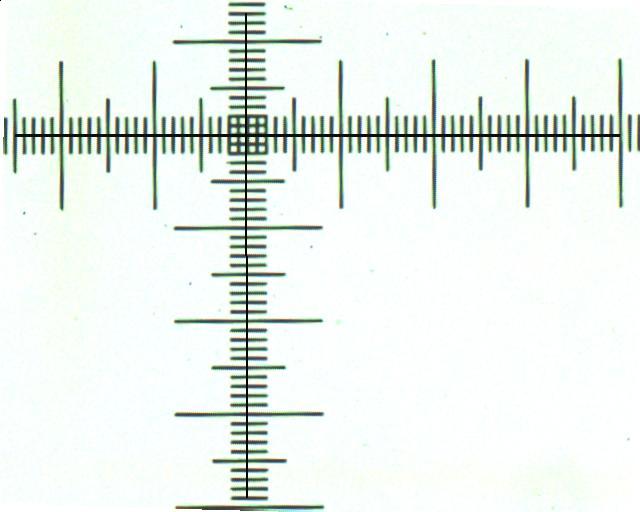
Magnification, Size, and Scale Bars

Measuring the size of Specimens viewed with a Microscope and adding Sc |  Motic Microscopes
Measuring the size of Specimens viewed with a Microscope and adding Sc | Motic Microscopes

How to set scale bar in imageJ on Microscope and camera images | image  calibration - YouTube
How to set scale bar in imageJ on Microscope and camera images | image calibration - YouTube

Measuring the size of Specimens viewed with a Microscope and adding Sc |  Motic Microscopes
Measuring the size of Specimens viewed with a Microscope and adding Sc | Motic Microscopes

Calculate the scale – Extreme macro and Paleontology

Transparent Scale Bar Png - Scale Bar Microscopy Magnification, Png  Download , Transparent Png Image - PNGitem
Transparent Scale Bar Png - Scale Bar Microscopy Magnification, Png Download , Transparent Png Image - PNGitem

Adding a Scale Bar onto a Microscopy Image using PowerPoint/ImageJ |  Drawing/Graphing-09 - YouTube
Adding a Scale Bar onto a Microscopy Image using PowerPoint/ImageJ | Drawing/Graphing-09 - YouTube